Upper Thigh Cross Sectional Anatomy - Thigh - Wikipedia : Prep for a quiz or learn for fun!. ;pocket atlas of sectional anatomy, computed tomography and magnetic resonance imaging, vol. Cross sectional anatomy, timothy f. Study cross sectional anatomy using smart web & mobile flashcards created by top students, teachers, and professors. Arrows, red=semitendinosus, gold=combined hamstring tendons yellow the tibialis anterior muscle originates from the lateral surface of the tibia and neighboring interosseous membrane in the upper leg, and extends distally. Axial slice of mri with all anatomical structures labeled.
;pocket atlas of sectional anatomy, computed tomography and magnetic resonance imaging, vol. Study cross sectional anatomy using smart web & mobile flashcards created by top students, teachers, and professors. Top cross sectional anatomy flashcards ranked by quality. An atlas of cross sectional human anatomy. Use the mouse scroll wheel to move the images up and down alternatively use the tiny arrows (>>) on both side of the image to move the images.
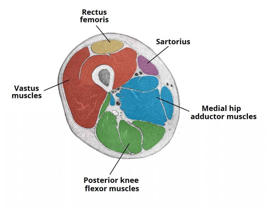
chest anatomy | MRI chest (thorax)axial anatomy | free cross sectional anatomy from mrimaster.com Cross sectional anatomy of the hip : The outer zone contains many myelinated axons that run up and down the spinal cord. This is mainly due to the fact that the three muscle compartments (figure 6) in the thigh can compensate much higher volumes than the four compartments below the knee 1. Study cross sectional anatomy using smart web & mobile flashcards created by top students, teachers, and professors. Head and neck thorax abdomen upper limbs lower limbs. See more ideas about anatomy, anatomy and physiology, medical anatomy. Computed tomography and magnetic resonance imaging. Surface anatomy is best studied using a regional.
It is typically used to describe the contraction properties of pennate muscles. Cross sectional anatomy, timothy f. The outer zone contains many myelinated axons that run up and down the spinal cord. This mri brain cross sectional anatomy tool is absolutely free to use. Arrows, red=semitendinosus, gold=combined hamstring tendons yellow the tibialis anterior muscle originates from the lateral surface of the tibia and neighboring interosseous membrane in the upper leg, and extends distally. Free online quiz thigh cross sectional anatomy practice. Approach the regions are as follows: ;pocket atlas of sectional anatomy, computed tomography and magnetic resonance imaging, vol. An atlas of cross sectional human anatomy. Surface anatomy is best studied using a regional. Dutra, human anatomy, anatomical sections, ct scan, computed axial tomography, mri scan, magnetic resonance imaging, virtual autopsy, physician, medical student, reference. Prep for a quiz or learn for fun! This article will describe classical cadaveric cross sections taken at various levels.
This is mainly due to the fact that the three muscle compartments (figure 6) in the thigh can compensate much higher volumes than the four compartments below the knee 1. Dutra, human anatomy, anatomical sections, ct scan, computed axial tomography, mri scan, magnetic resonance imaging, virtual autopsy, physician, medical student, reference. Cross sectional anatomy of the hip : The outer zone contains many myelinated axons that run up and down the spinal cord. Surface anatomy is best studied using a regional.
mri anatomy of elbow | axial cross sectional anatomy of elbow joint from mrimaster.com Femur pelvic girdle connective tissues that envelop the thigh: This article will describe classical cadaveric cross sections taken at various levels. This mri brain cross sectional anatomy tool is absolutely free to use. Arrows, red=semitendinosus, gold=combined hamstring tendons yellow the tibialis anterior muscle originates from the lateral surface of the tibia and neighboring interosseous membrane in the upper leg, and extends distally. Cross sectional anatomy of the hip : Free online quiz thigh cross sectional anatomy practice. This work provides the reader with a brief and easily. They divide the body into upper and lower portions, and like the sagittal and frontal planes, you need to have a reference point to know exactly where a.
These horizontal planes pass through the body at right angles to the midsagittal and the frontal planes.
• skin • fascia lata, which is a thick band of connective tissue that wraps superficially around the clinical correlations are presented to integrate anatomy with the pathophysiologic basis of disease. This is mainly due to the fact that the three muscle compartments (figure 6) in the thigh can compensate much higher volumes than the four compartments below the knee 1. Figure 3.4 major muscles of the upper extremities: Human sectional anatomy atlas of body sections, ct and mri images, fourth edition 4th edition 2015 unitedvrg.pdf. Dutra, human anatomy, anatomical sections, ct scan, computed axial tomography, mri scan, magnetic resonance imaging, virtual autopsy, physician, medical student, reference. The importance of sectional anatomy has already been explored in detail. Top cross sectional anatomy flashcards ranked by quality. Study cross sectional anatomy using smart web & mobile flashcards created by top students, teachers, and professors. Lecture presentation by steven bassett southeast community college. Mri of upper leg (femur). These horizontal planes pass through the body at right angles to the midsagittal and the frontal planes. Free online quiz thigh cross sectional anatomy practice. This mri brain cross sectional anatomy tool is absolutely free to use.
• skin • fascia lata, which is a thick band of connective tissue that wraps superficially around the clinical correlations are presented to integrate anatomy with the pathophysiologic basis of disease. The importance of sectional anatomy has already been explored in detail. Computed tomography and magnetic resonance imaging. Figure 3.4 major muscles of the upper extremities: Lecture presentation by steven bassett southeast community college.
Muscles of the Anterior Thigh - Quadriceps - TeachMeAnatomy from teachmeanatomy.info Prep for a quiz or learn for fun! This mri brain cross sectional anatomy tool is absolutely free to use. Figure 3.4 major muscles of the upper extremities: Study cross sectional anatomy using smart web & mobile flashcards created by top students, teachers, and professors. Use the mouse scroll wheel to move the images up and down alternatively use the tiny arrows (>>) on both side of the image to move the images. This article will describe classical cadaveric cross sections taken at various levels. Head and neck thorax abdomen upper limbs lower limbs. Computed tomography and magnetic resonance imaging.
This article will describe classical cadaveric cross sections taken at various levels. Femur pelvic girdle connective tissues that envelop the thigh: Cross sectional anatomy of the hip : Computed tomography and magnetic resonance imaging. Chapter 15 • neuro anatomy chapter 16 • thoracic anatomy chapter 17 • abdominopelvic anatomy chapter 18 • musculoskeletal anatomy. Surface anatomy is best studied using a regional. Free online quiz thigh cross sectional anatomy practice. An atlas of cross sectional human anatomy. Study cross sectional anatomy using smart web & mobile flashcards created by top students, teachers, and professors. • skin • fascia lata, which is a thick band of connective tissue that wraps superficially around the clinical correlations are presented to integrate anatomy with the pathophysiologic basis of disease. Data and dicom images (archived on a pacs (picture archiving and communicating system) were processed and exported as jpeg images. See more ideas about anatomy, anatomy and physiology, medical anatomy. They divide the body into upper and lower portions, and like the sagittal and frontal planes, you need to have a reference point to know exactly where a.
They divide the body into upper and lower portions, and like the sagittal and frontal planes, you need to have a reference point to know exactly where a upper thigh anatomy. Human sectional anatomy atlas of body sections, ct and mri images, fourth edition 4th edition 2015 unitedvrg.pdf.
Upper Thigh Cross Sectional Anatomy - Thigh - Wikipedia : Prep for a quiz or learn for fun!. There are any Upper Thigh Cross Sectional Anatomy - Thigh - Wikipedia : Prep for a quiz or learn for fun! in here.
%20axial%20image%2011.jpg)